facs flow cytometry protocol
Propidium iodide is a suspected carcinogen and should be handled with care. In general researchers will stain between 1 x 10 5 and 1 x 10 6 cells per sample.

Flow Cytometry Creative Biolabs
It is recommended that experimental conditions such as antibody concentration incubation time and temperature be optimized for each flow cytometry experiment.
. Download Flow Cytometry Protocols Handbook. 1 They can. Prepare your cell suspensions for Flow Cytometry.
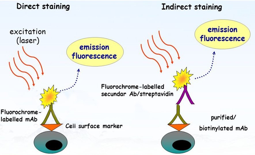
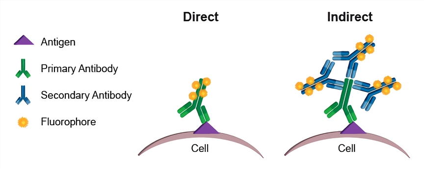
General procedure for flow cytometry using a conjugated primary antibody. Incubate for at least 30 min at room temperature or 4C in. Flow cytometry provides a rapid method to quantify cell characteristics.
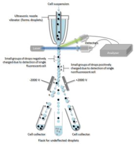
Print this indirect flow cytometry protocol. By utilizing highly specific antibodies labeled with fluorescent conjugates FACS analysis allows us to simultaneously collect data on and sort a biological sample by a nearly limitless number of. It provides a method for sorting a heterogeneous mixture of biological cells into two or more containers one cell at a time based upon the specific light scattering and fluorescent characteristics of each cell.
What is the purpose of flow cytometry. Introducing a combination of next-level spectral flow cytometry technologies and educational resources that offers an unmatched level of elegance and empowerment. FACS is an abbreviation for fluorescence-activated cell sorting which is a flow cytometry technique that further adds a degree of functionality.
A regular use of flow cytometers is the determination of the density of specific molecules on the surface of one or more cells in a population. Controls must also be evaluated alongside the experimental samples to insure the data is collected and interpreted correctly. Fluorescence-activated cell sorting FACS is a specialized type of flow cytometry.
Please read the following cell viability protocol in its entirety before beginning. Direct flow cytometry FACS protocol. If you are unable to immediately read your samples on a cytometer keep them shielded from light and in a refrigerator set at 4-8C.
Cells are usually stained in polystyrene round bottom 12 x 75 mm 2 Falcon tubes. Fluorophore and reagent selection guide for flow cytometry. Spectral flow cytometry assays and reagents.
Download our membrane staining summary. FACS of live cells. Bigfoot Spectral Cell Sorter.
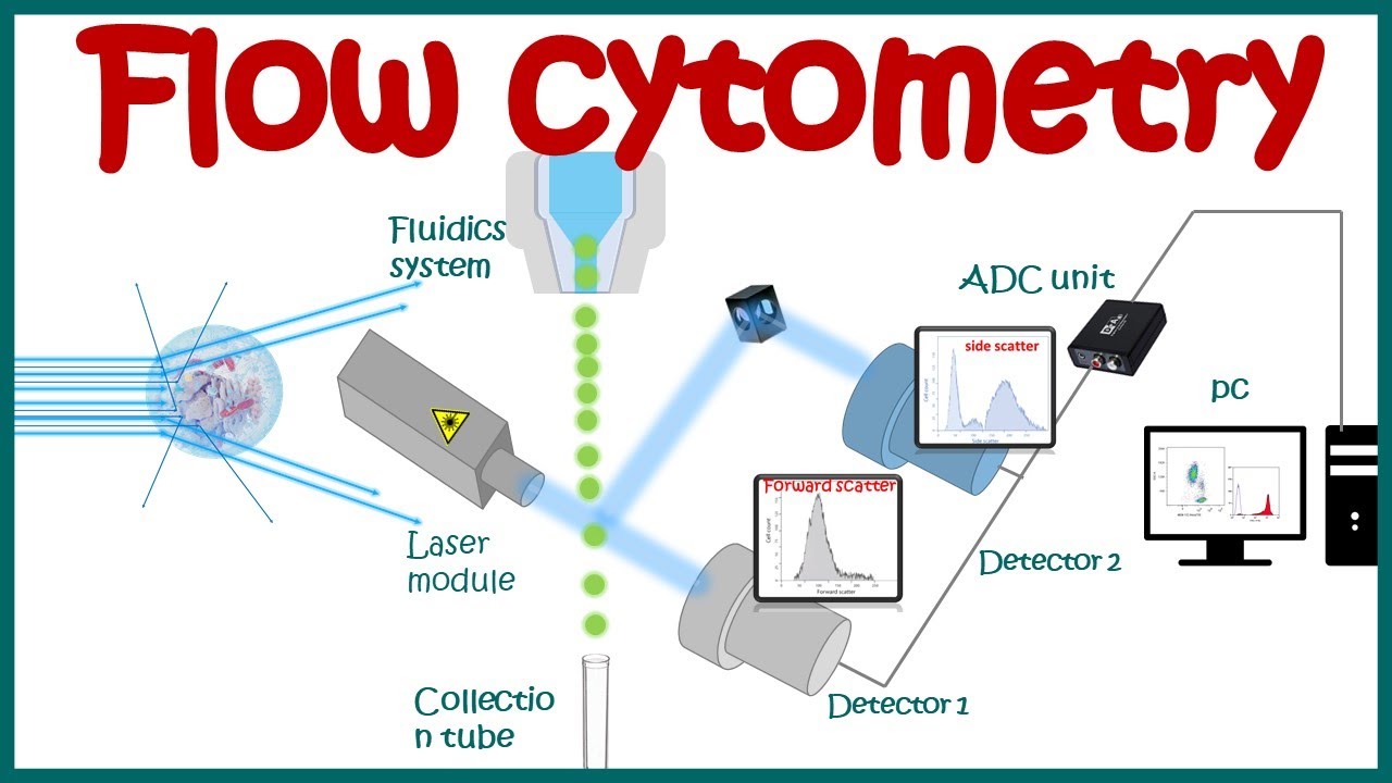
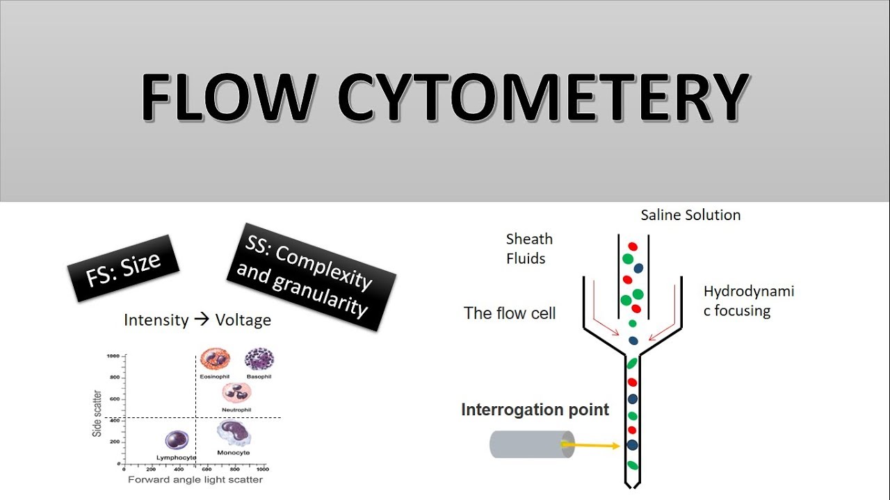
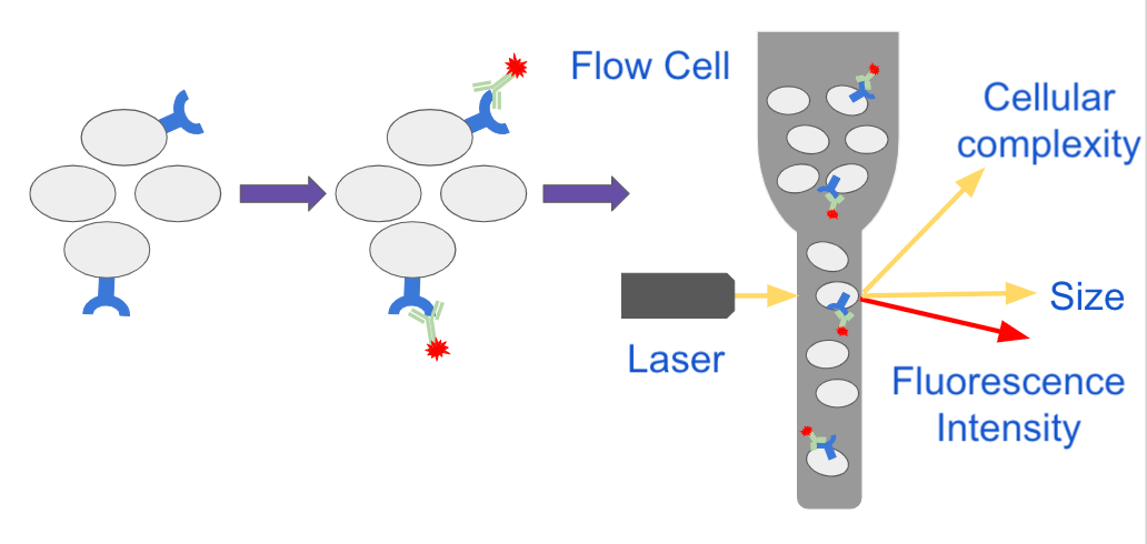
Since the tube uses spray-dried K2EDTA as opposed to liquid additives. From flow cytometers and sorters for simple to complex research applications to an extensive selection of reagents tools educational resources and protocols we support you in navigating your multicolor flow cytometry workflow journey. The properties measured include a particles relative size relative granularity or internal complexity and relative fluorescence intensity.
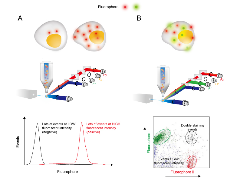
Sorting involves more complex mechanisms in the flow cytometer than a non-sorting analysis. Flow cytometry multicolor experiments may need compensation when there is fluorescence spillover Figure 1Pairing fluorochromes based on antigen density fluorochrome brightness and separating by channels helps to minimize the effects from spillover and may remove the need for compensation from smaller experiments. Spectral flow cytometry fundamentals.
Flow cytometry studies are used to identify and quantify immune cells and characterize hematological malignancies. As the number of antibodies used for phenotyping increases so does the complexity caused by the overlapping spectra of the fluorochromes. Incubate on ice for 20 min.
Invitrogen eBioscience ResourcesSelection guides Best Protocols product performance and more. The following protocol has been developed and optimized by RD Systems Flow Cytometry Laboratory for cell viability staining using propidium iodide. Spectral flow cytometry resources.
When the sample-filled tube is subjected to centrifugation the gel migrates and forms a physical barrier between the plasma and most of the cellular elements. The dye must be disposed of. However they can be stained in any container for which you have an.
Flow cytometry is a technology that simultaneously measures and then analyzes multiple physical characteristics of single particles usually cells as they flow in a fluid stream through a beam of light. Indirect labeling requires two incubation steps firstly with a primary antibody then with a compatible secondary antibody. General procedure for flow cytometry using a primary antibody and conjugated secondary antibody.
Intracellular Staining for Flow Cytometry How-To Videofor detecting cytokines and intranuclear markers. However as the number of parameters and colors. Flow cytometry multi-color selector.
Were stained with Invitrogen SYTO 9 nucleic acid stain and propidium iodide and then analyzed by flow cytometry according to the kit protocol. Harvest wash the cells and adjust cell suspension to a concentration of 1-5 x 10 6 cellsmL in ice-cold PBS 10 FCS 1 sodium azide. This is the basic task of flow cytometry.
Centrifuge at 1500 rpm for 5 min at 4C. Although most flow cytometry experiments involve labeling populations of cells that are relatively abundant the number of cells required will vary depending upon the rarity of your cells. FACS of live cells separates a population of cells into sub-populations based on fluorescent labeling.
This protocol is designed for staining of cell surface proteins. Add 01-10 μgml of the primary labeled antibody. The following flow cytometry staining protocol has been developed and optimized by RD Systems Flow Cytometry Laboratory.
The Intacellular Flow Cytometry Staining Protocol describes the process for intracellular staining of various cell types in vivo-stimulated tissues in vitro-stimulated cultures and whole blood for flow cytometry using BioLegends proprietary buffers and antibodies. However most flow cytometers cannot directly provide the cell concentration or absolute count of cells in a sample. PPT is an evacuated sterile blood collection tube that contains an inert gel and spray-dried K2EDTA anticoagulant for achieving plasma separation.
Fluorochrome chart a complete guide. Built on more than 45 years of BD experience and leadership in flow cytometry and multicolor analysis the BD FACSCanto II Flow Cytometry Systems deliver reliable performance accuracy and ease-of-use for todays busy clinical laboratories. Flow cytometry provides a well-established method to identify cells in solution and is most commonly used for evaluating peripheral blood bone marrow and other body fluids.
Flow Cytometry Fundamental Principle How FACS Works. The BD Accuri C6 Plus Flow Cytometer with its compact 11 x 1475 x 165-inch footprint light weight of 30 lb and operational simplicity supports a wide variety of applications including immunology cell and cancer biology plant and microbiology and industrial applications. Analysis of cell cycle.
Intracellular Staining Permeabilization Wash Buffer is used to permeabilize cells following fixation. Flow cytometry is a popular cell biology technique that utilizes laser-based technology to count sort and profile cells in a heterogeneous fluid mixture. Indirect flow cytometry FACS protocol.
These measurements may be relative semiquantitative or quantitative depending. Add 100 μl of Fc block to each sample Fc block diluted in FACS buffer at 150 ratio. Dilutions if necessary should be made in FACS buffer.
Spectral Flow Cytometry Fundamentals. Perform fluorescence activated cell sorting FACS or flow cytometric analysis. Get more information from the BD FACSCanto II System brochure.
The samples should be resuspended in Cell Staining Buffer.

In The Protocol Developed By Bernhard Fuchs S Team Bacterial Groups Are Enriched In Three Steps 1 In Situ Hybridization Postdoctoral Researcher Microbiology

Flow Cytometry Detection Of Surface And Intracellular Antigens In Pancreas From A Single Mouse Embryo Star Protocols

Flow Cytometry Based Protocols For Human Blood Marrow Immunophenotyping With Minimal Sample Perturbation Star Protocols
Quantitative Flow Cytometry Measurements Nist

Flow Cytometry Sample Preparation Proteintech Group
Direct Staining Flow Cytometry Creative Biolabs

Diagnostic Potential Of Imaging Flow Cytometry Trends In Biotechnology
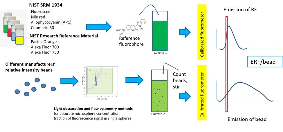
Quantitative Flow Cytometry Measurements Nist
Flow Cytometry Creative Biolabs

Flow Cytometry Facs Protocols Sino Biological

Flow Cytometry Introduction Abcam
Flow Cytometry Basic Principles What The Use Of Flow Cytometry Cell Sorting By Facs Youtube
The Principle Of Flow Cytometry And Facs 2 Facs Fluorescence Activated Cell Sorting Youtube
How Does Flow Cytometry Work Nanocellect

Single Cell Rna Expression Analysis Using Flow Cytometry Based On Specific Probe Ligation And Rolling Circle Amplification Acs Sensors

A Typical Flow Cytometry Experiment Sample Preparation From Blood Often Download Scientific Diagram
Flow Cytometry Guide Creative Diagnostics
Analyzing Single Cells With Flow Cytometry

Optimized Flow Cytometric Protocol For The Detection Of Functional Subsets Of Low Frequency Antigen Specific Cd4 And Cd8 T Cells Sciencedirect